Diagnostics
What can we do?
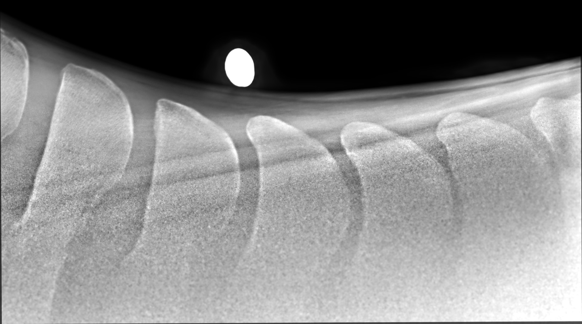
Diagnostics refer to the various methods used by veterinary surgeons to diagnose health problems in horses. These methods include physical examination, blood tests and imaging such as X-rays, ultrasound and endoscopy.
Ultrasonography
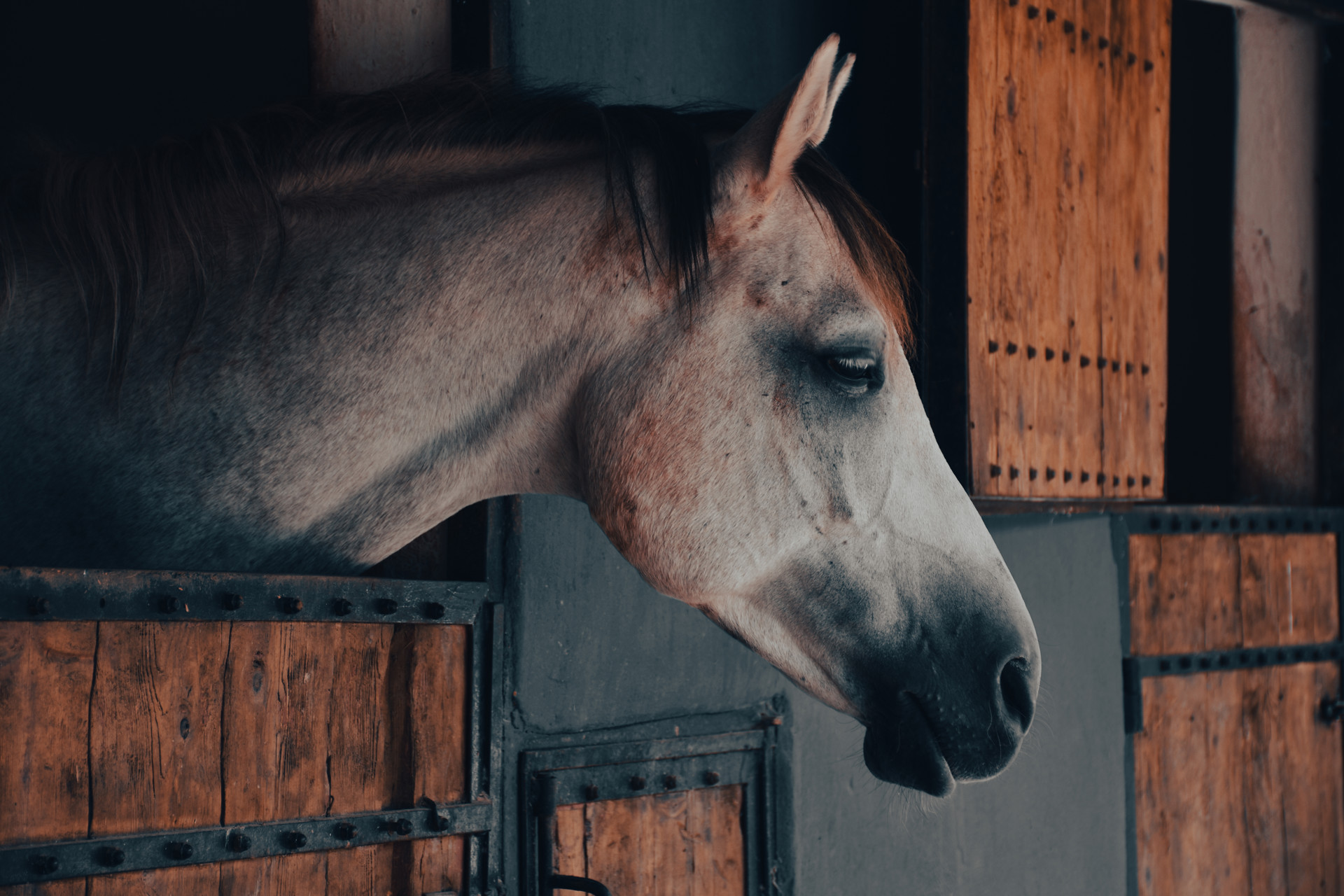
Our clinic has a wide range of ultrasound scanners, all portable, which are suitable for a variety of uses from scanning mares for pregnancy to diagnosing tendon injuries.
Ultrasound is primarily used for looking at soft-tissue structures, but can also be used to pick up subtle changes on the surface of bone in some cases. Our newest machine is capable of running off battery power, so even if there is no mains supply - no problem!
Mobile DR X-Ray Machine
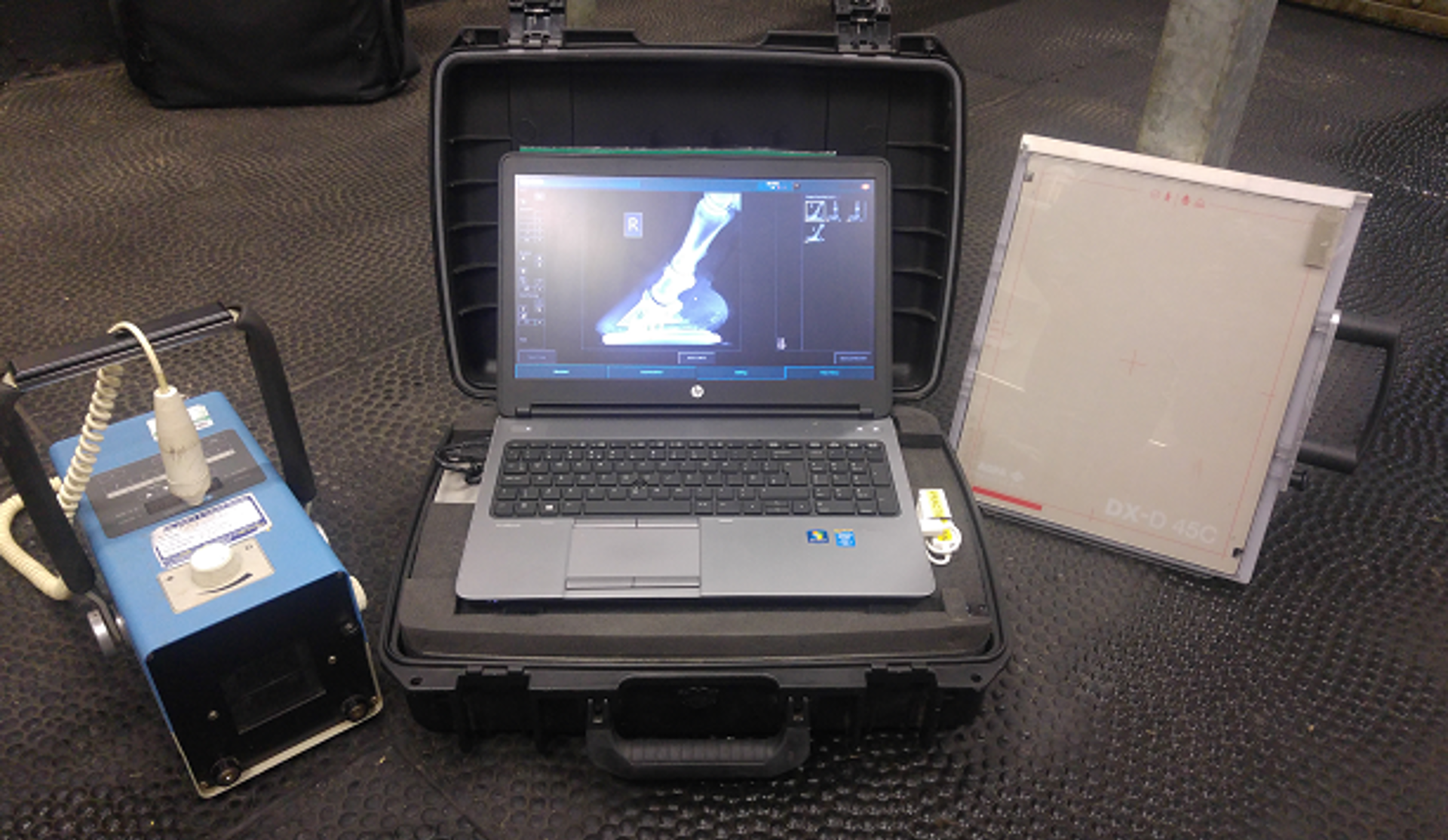
We always strive to offer the best service, investing in new technology to meet our customers’ needs. All of our clinics are equipped with mobile, wireless, DR X-ray systems. These systems have many benefits, including:
- Totally portable - X-rays can be taken at your premises; no need to transport your horse.
- Images can be displayed on the computer screen within seconds, so our vets can process them on site ensuring that we have the views we need before we leave your yard.
- Images are high resolution, with fantastic clarity.
- We can share the high resolution images with referral specialists where necessary.
- Wireless - no tripping over wires trailed across the yard or stable.
Endoscopy
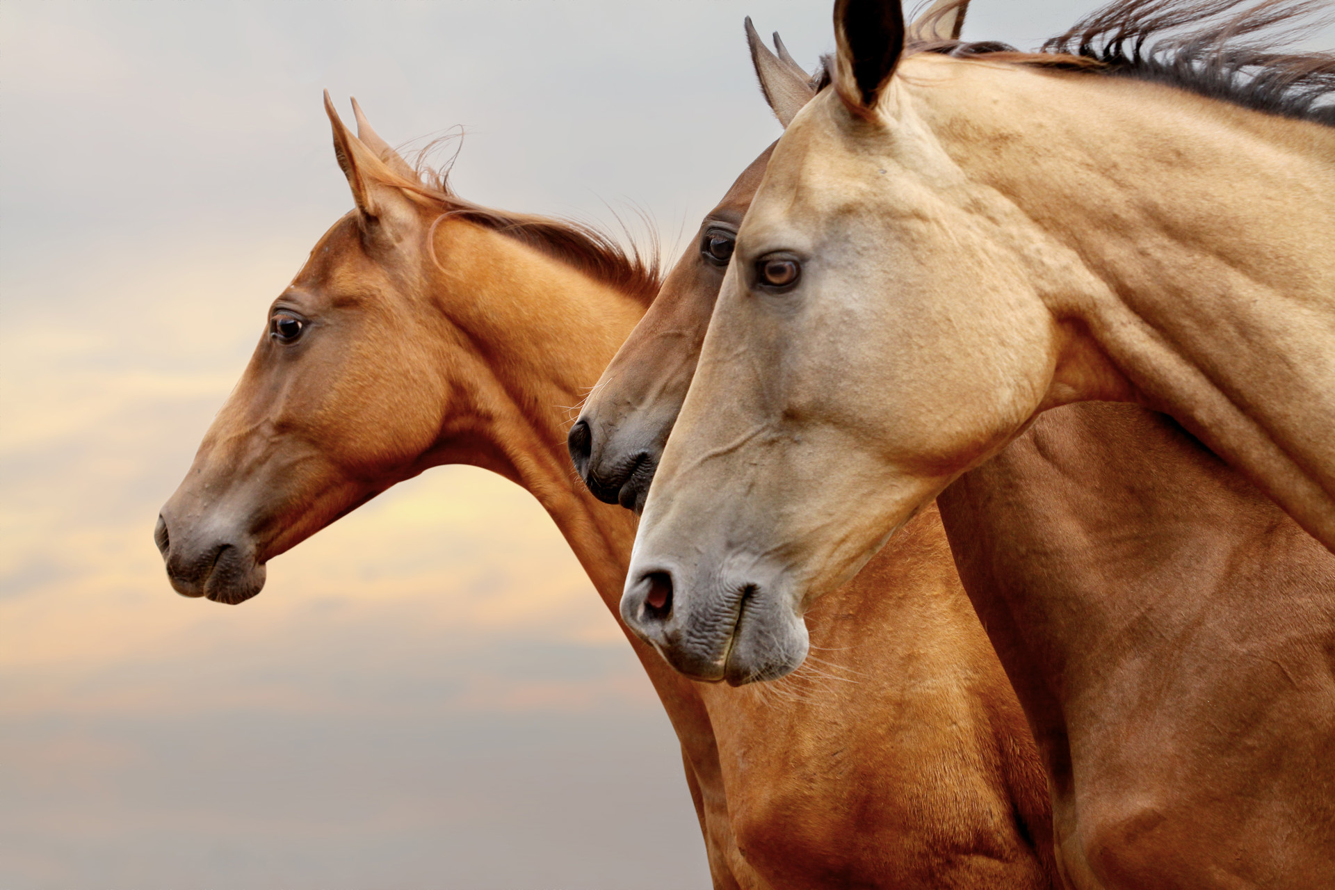
Used primarily to visualise the horse's throat and upper airways, this is an invaluable and portable tool to help in the diagnosis of respiratory disease.
Endoscopy can form an important part in the diagnosis process from subtle poor performance problems to overtly ill horses. An extra-long endoscope called a gastroscope is used to view the stomach and diagnose gastric ulcers.
MRI
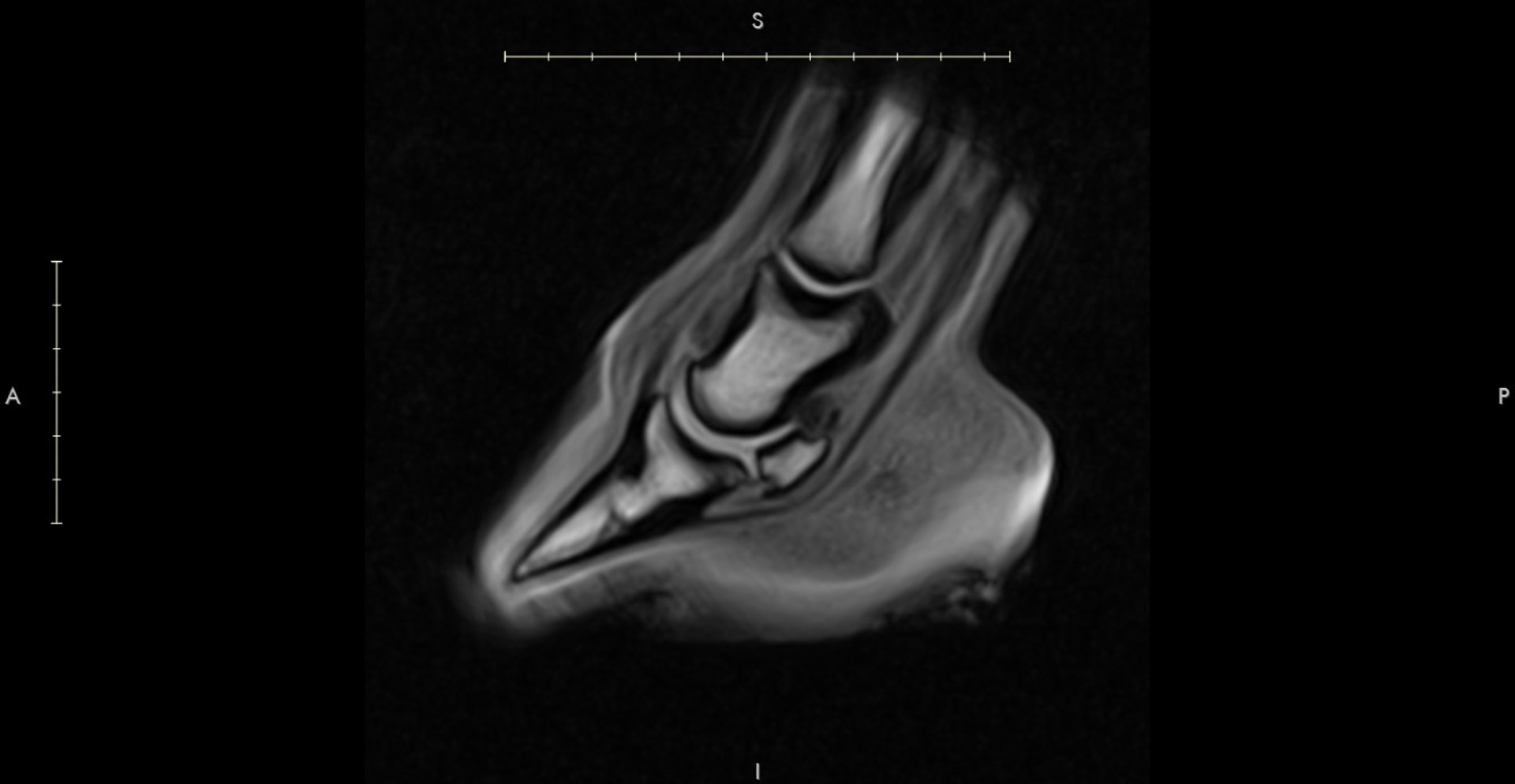
Magnetic Resonance Imaging (MRI) provides a detailed series of cross-sectional images of the soft tissues. If we decide that MRI is the best cause of action for your horse we would do this via referral.
MRI uses a powerful magnet, radio waves and a computer to create highly detailed images, it has become the “gold standard” for diagnosing some equine lameness, including those related to the foot and distal limb. Prior to an MRI scan your horse must have its shoes removed so that no metal enters the magnetic field.
CT scan
Computed tomography (CT) scanning is a non-invasive cross-sectional imaging technique that produces a 3D view of part of the body.
CT is performed in the standing sedated horse and can be especially useful for investigating poor performance, neck pain, disease in the head including sinus and nasal passage problems, trauma of the skull, headshaking and neurological diseases.
CT scanning is a specialist procedure and we would refer your horse if this was necessary.